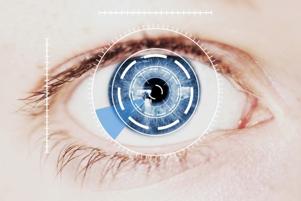
Early signs of many eye diseases may show up on the retina, or the back of the eye. retinal photography is a way of evaluating the back of the eye through color, high-resolution, digital photographs on which such signs may show up more easily.
Taking the images does not hurt at all. However, it does require the eyes to be dilated with special eye drops. After your eyes have been dilated, you will be sensitive to light for several hours. Because the photography takes place in a dark room, this is not bothersome until your appointment is over and you leave the office. You may need to wear dark glasses to shield your eyes from the light and have someone else drive you home. Eventually, the drugs that cause your eyes to dilate wear off and your pupils start reacting to light as usual.
Why is retinal photography performed?
As digital imaging of the retina becomes more advanced, efficient, and affordable, it may be performed more often. As of now, however, it is usually not performed as a part of routine eye exams. However, if your optometrist identifies symptoms of potential eye disease as part of routine screening, retinal photography may be ordered to get a clearer, more detailed image of the back of the eye and to confirm or rule out a diagnosis.
Digital imaging of the retina shows a number of important structures in the eye:
- Blood vessels that provide circulation to the eye
- The optic disk, where the optic nerve connects to the eye
- The macula, which allows the eye to focus and produce clear images
The following are eye conditions that retinal photography can help identify.
Glaucoma
Glaucoma occurs when the pressure inside the eye increases. This compresses the blood vessels in the eye and puts pressure on the optic nerve, resulting in vision loss.
Diabetic retinopathy
Diabetes can damage blood vessels in the eye, causing them to leak or swell. It can also cause new blood vessels to form. Early detection of diabetic retinopathy can prevent blindness through interventional treatment.
Macular degeneration
Most often associated with age, macular degeneration causes a loss of central vision because of changes to the blood vessels in the macula. With wet macular degeneration, which is the more severe type, the changes are due to the development of abnormal blood vessels in the macula. With dry macular degeneration, the existing blood vessels go through changes that cause them to become brittle and thin.
If you are diagnosed with an eye disease such as these, you may continue to have retinal photography performed at routine intervals to monitor your condition and further assess your condition. Even if you do not have an eye disease, photographing the retina offers the additional benefit of preserving a permanent record of changes in your eye over time.
Conclusion
Retinal photography can be performed to track changes in the eye and to diagnose ocular diseases. While the procedure itself is painless, it does require pupil dilation, which can cause discomfort for several hours thereafter until the medication wears off.
If you are experiencing any symptoms of possible eye disease or it has been a while since your last examination, contact our office to schedule an appointment.
Check out what others are saying about our services on Yelp: Retinal Photography in Dallas, TX.
Recent Posts
For those living with diabetes, undergoing a diabetic eye exam is one of the most important steps in protecting their vision and overall eye health. High blood sugar levels can lead to a range of complications, including conditions that damage the eyes over time. These exams help detect these issues before they become serious, allowing…
Contact lenses provide clear vision and convenience for individuals who prefer an alternative to eyeglasses. However, proper care and maintenance are essential to prevent infections, irritation, and eye damage. Neglecting hygiene practices can lead to serious eye conditions, including corneal ulcers and keratitis. Understanding how to clean, store, and handle contact lenses ensures long-term eye…
Maintaining eye health and preventing long-term issues is the result of consistent and quality vision care. Many people focus on overall wellness but may overlook daily habits that support healthy eyesight. However, taking simple steps each day can protect vision, reduce eye strain, and prevent future complications. By making eye health a priority, it is…
Prescription contacts provide vision correction, comfort, and convenience for those who do not want to wear glasses. However, caring for and wearing contacts takes some getting used to. Learning to insert, remove, and maintain them will help ensure a comfortable and safe experience.Not all contact lenses are the same, and choosing the right pair is…